Sunday, 26 December 2010

advanced elbow anatomy


descriptions

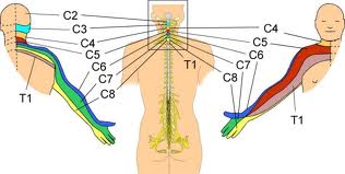
elbow neurology


elbow injuries

olecranon bursitis (students elbow)

lateral epicondylitis ( tennis elbow)

medial epicondylitis ( golfers/throwers elbow)

hyperextension injury

entrapment of radial nerve - tinels sign for radial nerve

entrapment of ulnar nerve - tinels sign for ulnar nerve

(entrapment of median nerve - phalens Test - can be reversed)

inflammation of biceps tendon

inflammation of triceps tendon

medial elbow ligament strain

elbow dislocation - radius/ulna both - 2nd most common after shoulder - posterior dislocation most common - associated with FOOSH - often associated with fracture - subluxation is small degree - complete dislocation requires manipulation


neurological examination - extended


look - inspect for tremors muscle twitches (fasciltations)
posture ( hypertonia UL flexion LL extension)

feel - muscle wastage

move


look for tone (smooth resistance to passive force)
power ( dominant side normally stronger )
coordination - rapid alternating movements
/ point to point - eyes open => cerebellar - eyes close => proproception
walking - hopping - narrow lunging
romberg - cerebellar/position sense
pronator drift - downward drifting ( mild hemiparesis - less severe than
hemiplegia)
sensation tests ( missing from main
reflexes - biceps reflexes triceps reflex briachoradialis
knee reflex ankle reflex ( babinski )

special tests

neurological examination - screen

3 minute

elbow normal range of motion

flexion 140-150

objective elbow exam

objective elbow exam

1. Working Hypothesis

2. Plan & Consent

3. Look :

3.1. General Obs

3.2. Acute Obs

4. Neurological screen

5. Feel - Palpation

6. Move :

6.1. A.P.R
A. In standing bilateral arms abducted 90 degrees ask to flex and extend
supination elbows flexed and touching side thumbs up
P omit if no problems in A.
R.


6.2. specialised tests
tinels sign


6.3. functional tests


7. Review